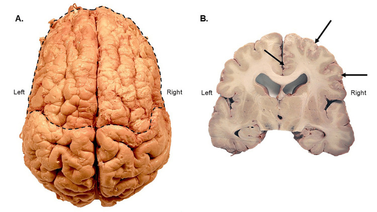
Figure 2. Superior and coronal views of the polymicrogyria (PMG) brain.
A. Superior view of the removed brain. Polymicrogyria (PMG) is evident in the outlined area when compared to the typical gyration of the more posterior brain. B. Coronal brain section at the level of the substantia nigra. The intact and fully developed corpus callosum is evident in this section; also of note is the stippled gray matter of the right hemisphere, as indicated by the arrows. Mild to moderate enlargement of the lateral ventricles was observed.

