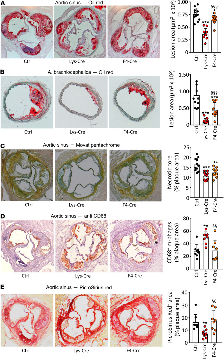
Figure 1. S1P1 overexpression in macrophages and monocytes retards atherosclerotic lesion development and alters plaque morphology in Ldlr–/– mice.
Aortic root (A and C–E) and brachiocephalic arteries (B) from WD-fed Ldlr–/– mice transplanted with S1pr1-KI (Ctrl, n = 11), S1pr1-LysMCre (Lys-Cre, n = 10), or S1pr1-F4/80Cre (F4-Cre, n = 10) BM were used for morphometry (A and B) or stained for necrotic core analysis (Movat pentachrome, C), macrophages (anti-CD68, D), or collagen (Picrosirius red, E). Bar graphs show the necrotic core extent or the macrophage or collagen content in plaques expressed as the percentage of lesion area. * - P < 0.05, ** - P < 0.01, *** - P < 0.001 (Lys-Cre vs. Ctrl or F4-Cre vs. Ctrl), §§ - P < 0.01, §§§ - P < 0.001 (Lys-Cre vs. F4-Cre, 1-way ANOVA except B: Kruskal-Wallis h test).

