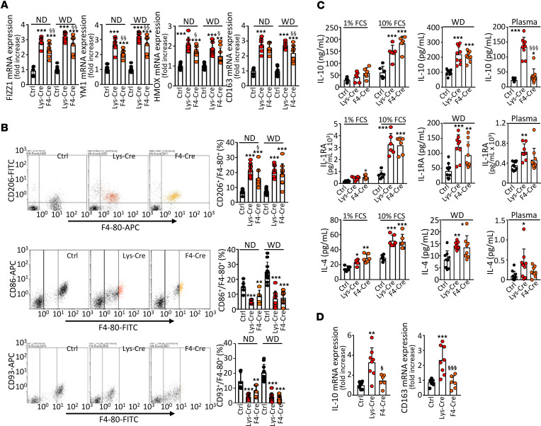
Figure 3. S1P1 overexpression in macrophages promotes antiinflammatory polarization.
PMs from either S1pr1-KI (Ctrl, n = 6–10), S1pr1-LysMCre (Lys-Cre, n = 6–10), or S1pr1-F4/80Cre (F4-Cre, n = 6–9) on ND or Ldlr–/– mice transplanted with S1pr1-KI (n = 9–11), S1pr1-LysMCre (n = 9–10), or S1pr1-F4/80Cre (n = 9–10) BM on WD. (A) qPCR of antiinflammatory signature genes. mRNA normalized to Gapdh and presented relative to S1pr1-KI. (B) CD206 (antiinflammatory marker) and CD86 and CD93 (pro-inflammatory markers) analyzed by flow cytometry. (C) PMs incubated for 24 hours in media containing 1% FCS (ND-fed mice, n = 6 for each group, left panels) or 10% FCS (ND- and WD-fed mice, left and central panels). Cytokines in media and plasmas (WD-fed mice, n = 8–11 for each group, central and right panels) determined by ELISA. (D) Cytokine mRNA expression in aortas by qPCR (n = 6–8 for each group). * - P < 0.05, ** - P < 0.01, *** - P < 0.001 (Lys-Cre vs. Ctrl or F4-Cre vs. Ctrl, § - P < 0.05, §§ - P < 0.01, §§§ - P < 0.001 (Lys-Cre vs. F4-Cre, 1-way ANOVA except C IL-10/Plasma and C IL-4/Plasma: Kruskal-Wallis h test).

