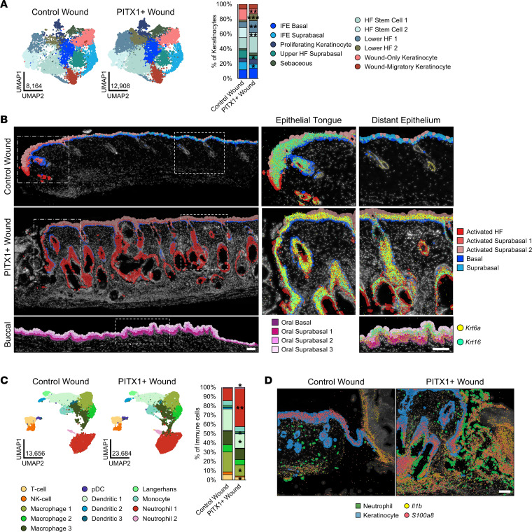
Figure 7. Primed, activated keratinocytes and enhanced neutrophil recruitment are features of PITX1+ skin wounds.
(A) UMAPs of control skin (n = 8), PITX1+ (n = 8), and buccal mucosa (n = 7) scRNA-Seq keratinocyte subpopulations (left). Proportion plot of wound keratinocyte subpopulations (right). (B) Representative Xenium of male control (n = 1) and PITX1+ wounds (n = 2) with cell subtypes highlighted. Dotted boxes indicate magnification of the wound-adjacent epithelial tongue (middle) and distal epithelium (right). Krt6a (yellow) and Krt16 (cyan) transcripts represented by colored dots. Scale bars of both large wound field and insets are 100 μm. (C) UMAPs of control and PITX1+ wound scRNA-Seq immune cell subpopulations (left). Proportion plot of immune cell subpopulations (right). pDC, plasmacytoid dendritic cell. (D) Representative Xenium of wound-adjacent region of male control and PITX1+ samples with Neutrophils (green) and Keratinocytes (blue) highlighted. Il1b (yellow) and S100a8 (red) transcripts represented by colored dots. Scale bar = 100 μm. Significance for proportion plots was assessed by proportionality testing followed by ad hoc comparisons against the corresponding cell type in control skin to derive log2 fold-change (log2FC) (*P < 0.01 & log2FC > |1.5|, **P < 0.01 & log2FC > |2|, ***P < 0.01 & log2FC > |4|).

