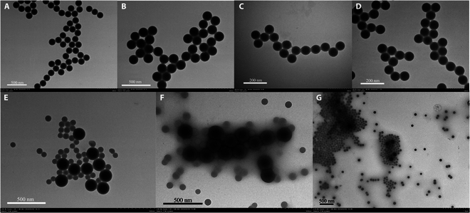
Fig. 4.

Results of latex spheres under different CRP states in transmission electron microscopy (TEM). (A) Large latex sphere bare balls. (B) Large immunolatex after coupling with antibodies. (C) Small latex sphere bare balls. (D) Small immunolatex after coupling with antibodies. (E) Mixture of immunolatex after coupling large and small latex spheres with antibodies. (F) Latex after reacting with low-concentration CRP. (G) Latex after reacting with high-concentration CRP.
