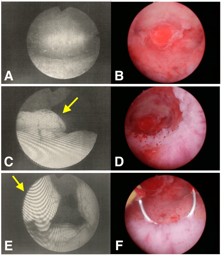
Figure 2. Hysteroscopic findings before and after administration of the GnRH antagonist.
The left column shows the hysteroscopic findings from a previous physician. Endometrial polyps with atypical vessels could be seen. The right column shows the observation during our hysteroscopy after GnRH antagonist administration. The lesions were broad-based, scar-like white, without abnormal blood vessels. A and B: Fundus of the uterus, no specific occupying lesions, floating desquamation is more evident under hysteroscopy during our procedure. C and D: Comparison in the visual field of polypoid tissue in the lower segment of the uterus, where atypical vessels were obscured and protruding polyps (yellow arrow) were absent after modification with the GnRH antagonist. E and F: The polypoid tissue has disappeared (yellow arrow), leaving white scarred tissue, and is excised through a trans-hysteroscopic energy device.
GnRH: Gonadotropin-releasing hormone

