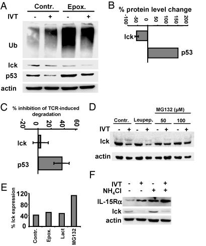
Fig. 3.
AID of lck is proteasome- and lysosome-independent but sensitive to inhibition by MG132. BK bulk CTLs were activated by IVT-pulsed APCs for 4 h in the absence (control) or presence of the indicated protease inhibitors. (A) Immunoblotting of lysates of BK289 cells with ubiquitin-, lck-, p53-, or actin-specific Abs. (B) Effect of epoxomycin on the steady-state levels of lck and p53. Intensities of lck- and p53-specific bands in lysates of cells incubated in the presence of epoxomycin were quantified and expressed as the percentage relative to control. Shown are the means ± SD of three experiments. (C) Effects of epoxomycin on AID of lck or p53 were analyzed and expressed as described in B. Shown are the means ± SD of three experiments. (D) Effect of MG132 on lck degradation assessed at the indicated concentrations. Cells activated by peptide-pulsed APCs in the presence of leupeptin were used as an additional control. (E) Quantification of the effect of MG132 at a concentration of 100 μM. Shown is one representative of three experiments in which samples of cells treated with lactacystin were also included. (F) Control or activated CTLs were cultured in the presence or absence of NH4Cl. Immunoblotting of total cell lysates with lck-, IL-15Rα-, or actin-specific Abs. Shown is one representative of five experiments.

