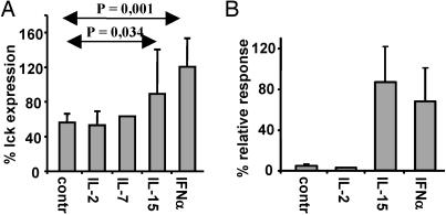
Fig. 7.
IL-15 and IFN-α increase lck expression after specific activation and restore responsiveness in preactivated CTLs. (A) The BK bulk CTLs were activated with peptide-pulsed APCs and cultured for 48 h in standard medium alone (control) or with the addition of exogenous IL-2, IL-7, IL-15, or IFN-α. The expression of lck was analyzed as described above. (B) Capacity of CTLs to proliferate in response to the secondary challenge as evaluated by a [3H]thymidine incorporation assay. The results are expressed as the percentage relative to thymidine incorporation of control CTLs, which were not preactivated with peptide-pulsed APCs and cultured in the absence (control) or presence of the indicated lymphokine. Shown are the means ± SD of four experiments.

