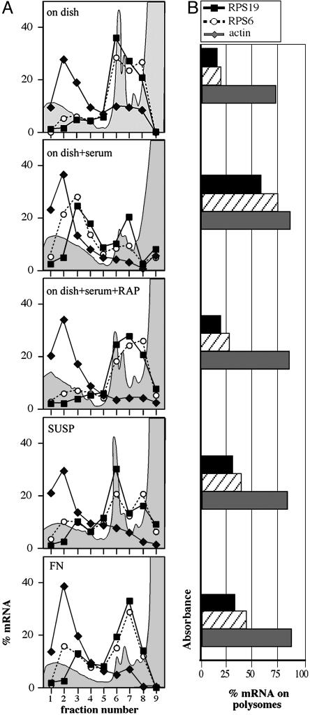
Fig. 4.
5′TOP mRNA translation is not stimulated by adhesion to FN. Northern blot was carried out with polysomal RNA from NIH 3T3 cells on plastic (C) after stimulation with serum (FBS) alone or with rapamycin (FBS+RAP), cells deprived of anchorage (SUSP) or spreading onto FN. 5′TOP mRNA probes (RPS19 and RPS6) and non-5′TOP mRNA probe as a control (β-actin) were used. Quantitation of the signal is reported as linear plot of the percentage of mRNA in each fraction (A) and as bar plot of the percentage of mRNA on polysomes (B), obtained by adding up the values of fractions 1-5. The absorbance profile is outlined (gray area) in the background of each plot.

