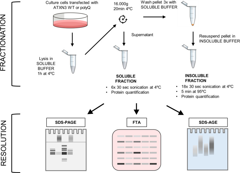
Fig 1. Experimental workflow of the fractionation assay.
Cultured cells expressing ATXN3 proteins are washed in PBS and lysed in soluble buffer. After centrifugation, the supernatant is saved as the soluble fraction. Pellets are washed with soluble buffer and resuspended with insoluble buffer, hereafter referred as the insoluble fraction. Fractions are sonicated, incubated for 5 min at 95°C for insoluble fractions, before quantification. Samples are then analysed by Polyacrylamide Gel Electrophoresis (SDS-PAGE), Filter Trap Assay (FTA) and SDS-Agarose Gel Electrophoresis (SDS-AGE) with the optimized parameters described in this article, to allow an optimal resolution and quantification depending on the ATXN3 species of interest.

