Fig. 2.
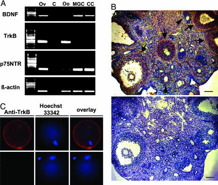
Localization of BDNF, TrkB, and p75 NTR transcripts as well as BDNF and TrkB antigens in the mouse ovary. (A) Expression of BDNF, TrkB, and p75 NTR mRNAs in isolated ovarian cells obtained from mice 48 h after PMSG treatment was detected by using nested RT-PCR. Levels of β-actin serve as loading controls. Total ovarian cDNA was used in positive control tests, whereas no template DNA was included for negative controls. Ov, ovary; C, negative control; Oo, oocyte; MGC, mural granulosa cells; CC, cumulus cells. (B) Immunohistochemical detection of BDNF in ovaries of PMSG-primed mice 7 h after hCG injection. BDNF was found in cumulus and mural granulosa cells of preovulatory follicles (arrows), whereas weaker staining was found in mural granulosa cells of small antral follicles (arrowheads). (Upper) BDNF staining. (Lower) Negative control. (Scale bars, 40 μm.) (C) Immunofluorescence staining of TrkB in an oocyte at the metaphase I stage. (Upper) TrkB staining. (Lower) Negative control. Overlay pictures showed combined staining of plasma membrane-bound TrkB and nuclear DNA (Hoechst staining).

