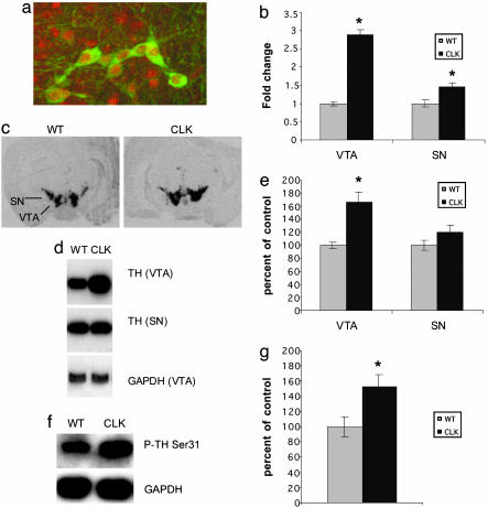
Fig. 3.
CLOCK is expressed in dopamine neurons, and TH levels and phosphorylation are increased in Clock mutants. (a) Sections containing the VTA were labeled with antibodies against CLOCK (red) and TH (green). Fluorescence was integrated by using confocal microscopy. Results are representative of multiple sections obtained throughout the anterior–posterior axis of the VTA of five mice (data not shown). (b–g) Clock mutants have increased TH protein and mRNA levels in the VTA. mRNA levels were determined from VTA and substantia nigra (SN) tissue punches from Clock mutants (Clk) and wild-type (WT) controls by real-time PCR (n = 5) (b) and in situ hybridization (n = 8). (c) Protein levels and phosphorylation (d–g) were determined by Western blot analysis (representative blots are shown in d and f, n = 5). In all cases, GAPDH was used as a control. *, P < 0.05 by ANOVA.

