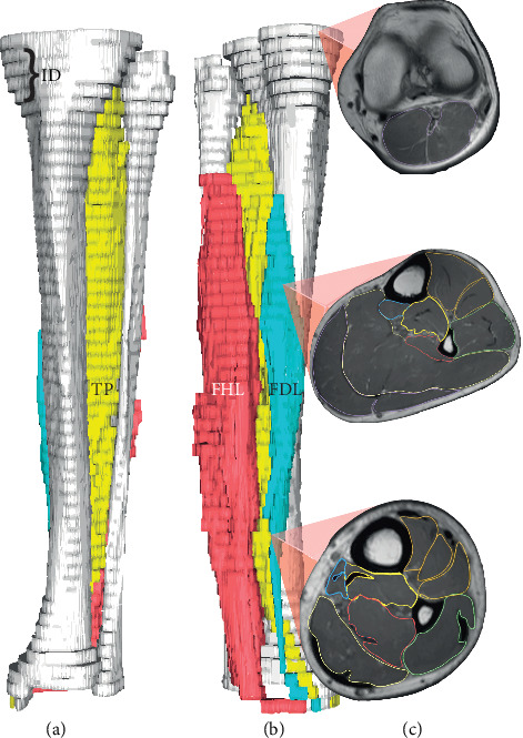
Figure 1.

3-D magnetic resonance rendered image and segmented slices of a MTSS group participant. (a) Anterior view, (b) posteromedial view, and (c) slice axial cross-sectional areas. ID, interslice space of 2.4 cm; FHL, flexor hallucis longus; FDL, flexor digitorum longus; TP, tibialis posterior.
