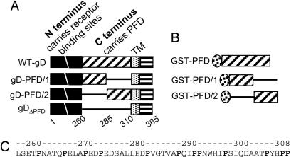
Fig. 1.
Schematic diagram of gD (A) and GST-PFD (B) constructs and PFD sequence (C). (A) Top line, linear map of WT-gD, with N terminus (amino acids 1-260) carrying receptor-binding sites (black) and C terminus (amino acids 260-310) carrying the PFD (diagonal lines), the transmembrane (TM) (dotted), and the C-tail (horizontal lines) regions. The regions are not shown to scale. The numbers indicate the region coordinates. gDΔPFD carries gD amino acids 1-260 and 50 aa from CD8, derived from the sequence upstream of TM (defined as CD8 amino acids 261-310), which replace gD amino acids 261-310. gD-PFD/1 carries gD amino acids 1-285 and CD8 amino acids 285-310. gD-PFD/2 carries gD amino acids 1-260 and 285-310 and CD8 amino acids 261-285. In all constructs, the TM and C-tail are from gD. (B) GST-PFD carries GST fused to gD amino acids 260-310. GST-PFD/1 carries GST fused to a segment composed of gD amino acids 260-285 and CD8 amino acids 285-310. GST-PFD/2 carries GST fused to a segment composed of CD8 amino acids 260-285 and gD amino acids 285-310. (C) PFD sequence and its coordinates.

