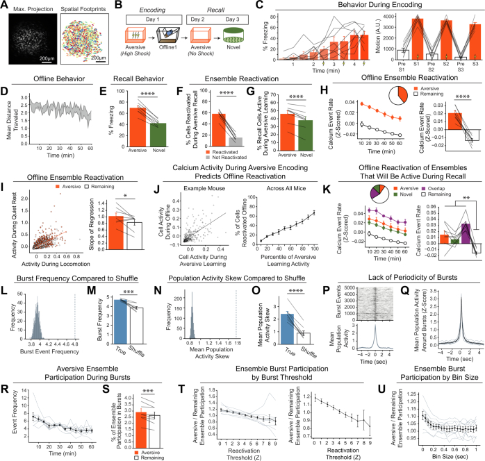
Extended Data Fig. 3. Neurons active during Aversive encoding are selectively reactivated offline and during Aversive recall.
A) Representative maximum intensity projection of the field-of-view of one example session (left). Spatial footprints of all recorded cells during the session, randomly colour-coded (right). B) Schematic of a single aversive experience. Mice had an Aversive experience followed by a 1 hr offline session in the homecage. The next day, mice were tested in the Aversive context, followed by a test in a Novel context one day later. Calcium imaging in hippocampal CA1 was performed during all sessions. C) Mice acquired within-session freezing during Aversive encoding (left); main effect of time (F8,56 = 12.59, p = 3.87e-10, n = 8 mice). And mice responded robustly to all three foot shocks, though their locomotion generally decreased across shocks, driven by increased freezing (right); main effect of shock number (F2,14 = 7.45, p = 0.0154, n = 8 mice) and main effect of PreShock vs Shock (F1,7 = 581, p = 5.38e-8, n = 8 mice), and no interaction. D) Mice displayed a modest decrease in locomotion across the 1 hr offline period (arbitrary units) (R2 = 0.064, p = 1.9e-8, n = 8 mice). E) Mice froze significantly more in the Aversive context than in a Novel context during recall (t7 = 165, p = 4e-6, n = 8 mice). F) Cells that were active during Aversive encoding and reactivated offline were significantly more likely to be reactivated during Aversive recall than cells active during Aversive encoding and not reactivated offline (t7 = 19.41, p = 2e-7, n = 8 mice). G) A larger fraction of cells active during Aversive recall than during Novel recall were previously active during Aversive encoding (t7 = 6.897, p = 0.0002, n = 8 mice). H) During the offline period, ~40% of the population was made up of cells previously active during Aversive encoding (top). This Aversive ensemble was much more highly active than the rest of the population during the offline period (bottom; A.U.) (t7 = 8.538, p = 0.00006, n = 8 mice). I) Each cell’s activity was compared during locomotion vs during quiet rest (left; A.U.). A regression line was fit to the cells in the Aversive ensemble and in the Remaining ensemble separately, for each mouse. The Remaining ensemble showed greater activity during locomotion than during quiet rest (i.e., a less positive slope). The Aversive ensemble showed relatively greater activity during quiet rest than locomotion (i.e., a more positive slope) across mice (right) (t7 = 5.76, p = 0.047, n = 8 mice). J) Cells that had high levels of activity (A.U.) during Aversive encoding continued to have high levels of activity during the offline period (example mouse; left). There was a linear relationship between how active a cell was during Aversive encoding and how likely it was to be reactivated during the offline period (all mice; right) (R2 = 0.726, p = 1.25e-23, n = 8 mice). K) During the offline period, cells that would go on to become active during recall were more highly active than the Remaining ensemble during the offline period. The top represents the proportion of each ensemble (legend to its right). The cells that would become active during both Aversive and Novel recall were most highly active (A.U.). There was no difference in activity in the cells that would go on to be active in Aversive or Novel. Main effect of Ensemble (F3,21 = 27.81, p = 1.65e-7, n = 8 mice). Post-hoc tests: for Aversive vs Novel (t7 = 1.33, p = 0.22), for Remaining vs Aversive ∩ Novel (t7 = 11.95, p = 0.000007), for Remaining vs Aversive (t7 = 3.97, p = 0.005), for Remaining vs Novel (t7 = 7.47, p = 0.0001). L) Neuron activities were circularly shuffled 1000 times relative to one another and the mean population activity was re-computed each time. This shuffling method preserved the autocorrelations for each neuron while disrupting the co-firing relationships between neurons. The burst frequency was computed for each of these shuffles to produce a shuffled burst frequency distribution (grey histogram), to which the true burst frequency was compared (blue dotted line). This is an example mouse. M) The mean burst frequency for the shuffled distribution was computed and compared to the true burst frequency for each mouse. True burst frequencies were greater than shuffled burst frequencies in every mouse (t7 = 6.159, p = 0.000463, n = 8 mice), suggesting that during the offline period, hippocampal CA1 neurons fire in a more coordinated manner than would be expected from shuffled neuronal activities. N) As in Extended Data Fig. 3l, neuron activities were shuffled, and mean population was re-computed each time. From this population activity trace, the skew of the distribution was computed. If there were distinct periods where many neurons simultaneously fired, we hypothesized that the true distribution of mean population activity would be more skewed with a strong right tail demonstrating large and brief deflections, compared to shuffled neuronal activities. We computed the skew of each shuffled mean population activity, to produce a distribution (grey histogram), to which the true mean population’s skew was compared (blue dotted line). This is an example mouse. O) The mean skew for the shuffled distribution was computed and compared to the true skew of the mean population activity for each mouse. The true skew was greater than the shuffled skew in every mouse (t7 = 13.36, p = 0.000003, n = 8 mice), supporting the idea that the mean population activity undergoes brief burst-like activations requiring the coordinated activity of groups of neurons. P) Matrix of burst events for an example mouse, stacked along the y-axis and centred on time t = 0 (top), and the average mean population activity around each burst event (bottom). Q) As in Extended Data Fig. 3p but averaged across all mice. Each thin line represents one mouse, and the thick black line represents the mean across mice with the grey ribbon around it representing the standard error (n = 8 mice). There is no periodicity to when these burst events occur. R) The burst event frequency decreased across the hour (F11,77 = 6.91, p = 5.66e-8, n = 8 mice). S) A larger fraction of the Aversive ensemble vs the Remaining ensemble participated in each burst event (left) (t7 = 3.68, p = 0.0079, n = 8 mice). T) Ensemble burst participation as a function of burst threshold. The burst threshold was parametrically varied, and the ratio of Aversive-to-Remaining burst participation was computed at each burst threshold. Aversive-to-Remaining burst ratio is negatively related to burst threshold (R2 = 0.28, p = 3.4e-7) (n = 8 mice). On the left graph, the black line represents the mean across mice with SEM represented in the error bars, and each individual mouse is represented by the grey lines. On the right is the same data as on the left graph, but without the individual mice. U) Ensemble burst participation as a function of bin size. The Aversive-to-Remaining burst participation ratio was computed at varying bin sizes. At larger bin sizes, the selective increase in Aversive burst participation is no longer present (n = 8 mice).

