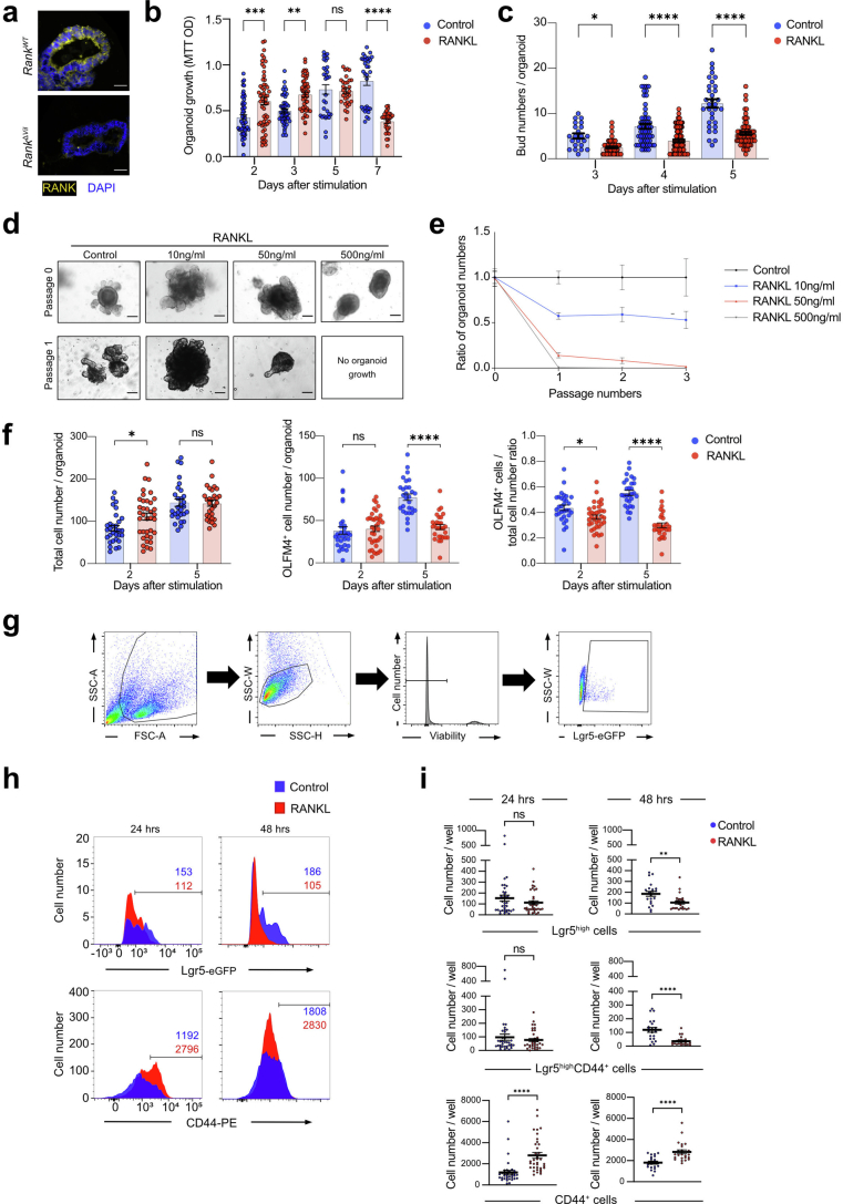
Extended Data Fig. 1. Characterization of stem cells in RANK–RANKL-stimulated mouse intestinal organoids.
a, Representative images of anti-RANK immunostaining in mouse jejunal intestinal organoids from RankWT and RankΔVil mice. DAPI is shown to visualize nuclei. Scale bars, 50 μm. b, Proliferation assay in organoids without (control) or in the presence of recombinant mouse RANKL (rmRANKL; 50 ng/ml) as determined by an MTT assay at the indicated time points. Each plot represents MTT OD, pooled from at least two independent experiments. Group numbers at 2 days: n = 56 (control), n = 56 (rmRANKL); 3 days: n = 50 (control), n = 50 (rmRANKL); 5 days: n = 31 (control), n = 31 (rmRANKL) and 7 days: n = 32 (control), n = 31 (rmRANKL). c, Numbers of buds per intestinal organoid cultured in ENR medium with/without rmRANKL. Data were combined from three independent experiments. Group numbers at 3 days: n = 22 (control), n = 59 (rmRANKL); 4 days: n = 57 (control), n = 102 (rmRANKL) and 5 days: n = 32 (control), n = 66 (rmRANKL) per group. d, Representative images of jejunal organoids at passage 0 and passage 1 cultured in the absence (control) or presence of the indicated concentrations of rmRANKL. Scale bars, 100 μm. e, Ratios of organoid numbers after prolonged passaging in the absence (control) and presence of rmRANKL. Numbers of organoids were counted at each passage. Data from two independent experiments are shown. n = 8 (control), n = 8 (10 ng/ml RANKL), n = 8 (50 ng/ml RANKL), n = 8 (500 ng/ml RANKL). f, Total cell numbers, OLFM4 positive cells per organoid and ratios of OLFM4 positive cell in relation to the total cell number per each jejunal organoid cultured in ENR medium with/without rmRANKL. Data were combined from two independent experiments. 2 days: n = 31 (control), n = 37 (rmRANKL); 5 days: n = 29 (control), n = 29 (rmRANKL) per group. g, Gating strategy for detecting Lgr5-eGFP+ cells using fluorescence-activated cell sorting (FACS). Viability was determined using the viability-dye described in the Methods. FSC, forward scatter; SSC, side scatter. h, Representative FACS histograms of Lgr5high cells and CD44+ cells isolated from Lgr5-eGFPiresCreER/+ jejunal organoids cultured without (control) and with rmRANKL (50 ng/ml) for the indicated times. Numbers of Lgr5high cells and CD44+ cells among total viable organoid cells are indicated for each group. Data are representative of at least two independent experiments. i, Quantification of Lgr5high, Lgr5highCD44+, and CD44+ cells per well in Lgr5-eGFPiresCreER/+ jejunal organoids cultured without (control) or with rmRANKL (50 ng/ml) for the indicated times. n = 36 (Control, 24hrs), n = 36 (RANKL, 24hrs), n = 21 (Control, 48hrs), n = 24 (RANKL, 48hrs). Data are mean ± s.e.m. *P < 0.05; **P < 0.01; ***P < 0.001; ****P < 0.0001; ns, not significant. One-way analysis of variance (ANOVA) with Tukey’s post hoc test (b,c,f); Two-tailed Student’s t-test (i). More details on statistics and reproducibility can be found in the Methods.

