
FIGURE 1.
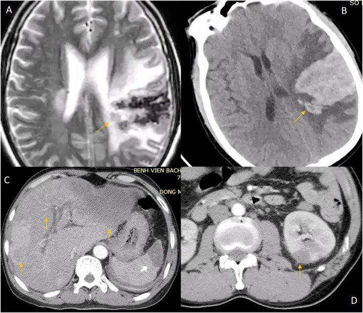
Extracardiac manifestations. (A) T2 weighted magnetic resonance imaging showing cerebral infarction with hemorrhagic transformation. (B) The same lesion on non‐contrast CT. Abdominal multi‐slice computed tomography (arterial phase) image showing: (C) Hypointense masses in left and right lobes of the liver (yellow arrows) and spleen 535 (white arrow). (D) Hypointense mass in the left kidney.