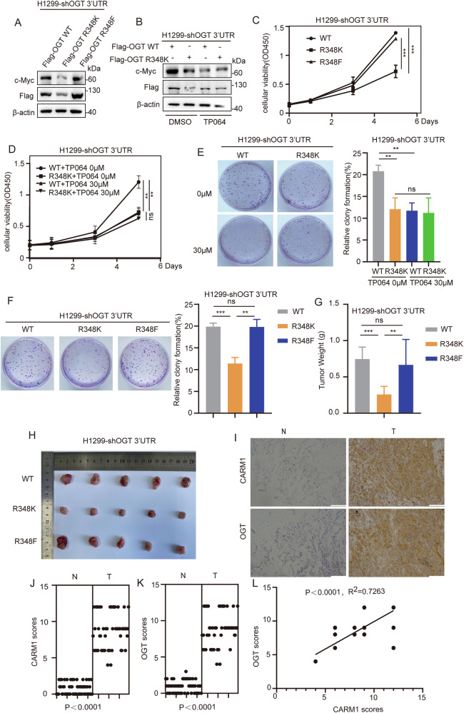
Fig. 6. Arginine methylation of OGT at R348 promotes NSCLC proliferation.
A H1299 cells were transfected with Flag-OGT WT /R348K/R348F after knockdown of endogenous OGT. The level of c-Myc in H1299 cells was assessed by western blotting. B The level of c-Myc was measured in H1299 cells re-expressing OGT WT/R348K after knockdown of endogenous OGT and then treated with TP064 for 24 h. c-Myc expression was assessed by western blotting. C, F H1299 cells were transfected with Flag-OGT WT/R348K/R348F after knockdown of endogenous OGT. Representative images of CCK8 (C) and colony formation (F) are shown. All data points are included (error bars represent the mean ± SD, n = 3 experimental replicates, **P < 0.01, ***P < 0.001, ns = not significant, Student’s t test). D, E H1299 cells expressing OGT WT/R348K after knockdown of endogenous OGT were treated with TP064 for 24 h. Representative images of CCK8 (D) and colony formation (E) are shown. All data points are included (error bars represent the mean ± SD, n = 3 experimental replicates, **P < 0.01, ns = not significant, Student’s t test). G, H H1299-shOGT-WT/R348K/R348F cells (3 × 10⁶) were injected into the skin of 5-week-old-male nude mice, and 3 weeks later, mice were sacrificed by euthanasia. Tumor size (H) and weight (G) were measured (error bars indicate the mean ± SD, n = 5 mice for each group, ns=not significant, **P < 0.01, ***P < 0.001, Student’s t test). I Representative images of immunohistochemical staining of OGT and CARM1 in human non-small cell lung cancer samples. Scale bars, 100 μm. J,K The expression levels of OGT (K) and CARM1 (J) in human non-small cell lung cancer tissues and adjacent tissues were measured (n = 59 non-small cell lung tumor samples, P < 0.0001, Student’s t test). L The correlation of OGT with CARM1 was statistically significant in human non-small cell lung cancer samples (n = 59 human non-small cell lung cancer samples; R2 = 0.7263, P < 0.0001; Pearson’s correlation test). The immunohistochemistry score was calculated according to Feng et al. [26].

