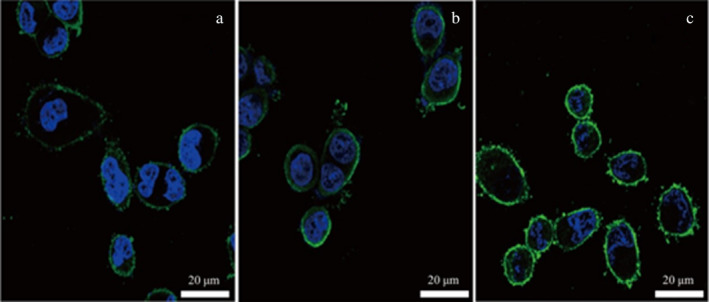
FIGURE 8.

Adhesion of different concentrations of surface protein to chicken intestinal epithelial cells. (a) Fluorescence microscope combined image of low concentration of surface protein adhering to chicken intestinal epithelial mucosa cells. (b) Fluorescence microscope combined image of medium concentration of surface protein adhering to chicken intestinal epithelial mucosa cells. (c) Fluorescence microscope combined image of high concentration of surface protein adhering to chicken intestinal epithelial mucosa cells.
