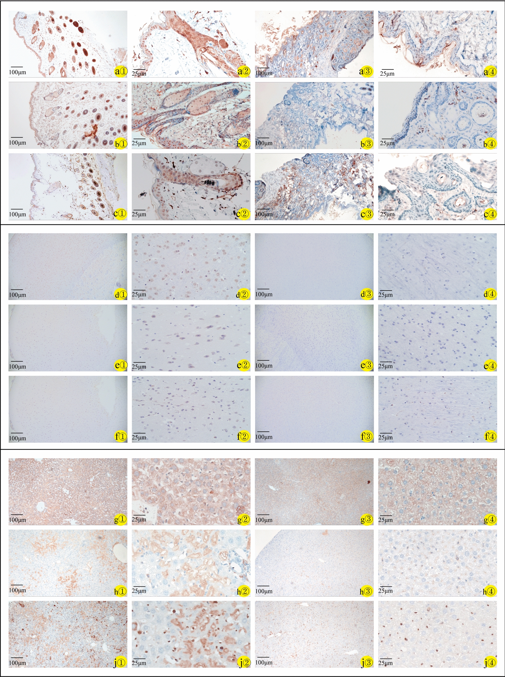
Fig. 6.

Immunohistochemistry of skin, brain, and liver specimens of wild-type mice (a①–j① × 100; a②–j② × 400) and NCSTN gene knockout mice (a③–j③ × 100; a④–j④ × 400). Staining for nicastrin (WT-a①/a② and NCSTN gene knockout mice-a③/a④), NICD1 (WT-b①/b② and NCSTN gene knockout mice-b③/b④), and hes1(WT-c①/c② and NCSTN gene knockout mice-c③/c④) in skin tissues mainly in epidermis and hair follicles; in brain tissues, staining for nicastrin (WT-d①/d② and NCSTN gene knockout mice-d③/d④) mainly in neuronal cells and glial nuclear plasma. NICD1 (WT-e①/e② and NCSTN gene knockout mice-e③/e④) mainly expressed in glial nuclear plasma, with a small amount in nerve cells, and hes1(WT-f①/f② and NCSTN gene knockout mice-f③/f④) expressed in neuronal cells, with a small amount in glial nuclear plasma. In liver tissues, staining for nicastrin (WT-g①/g② and NCSTN gene knockout mice-g③/g④), NICD1 (WT-h①/h② and NCSTN gene knockout mice-h③/h④), and hes1(WT-j①/j② and NCSTN gene knockout mice-j③/j④) mainly in the nucleus of hepatic sinusoidal endothelial cells. Compared with wild-type mice (a①–j①, a②–j②), the expression of these proteins in skin, brain and liver tissue was lower in NCSTN gene knockout mice (a③–j③, a④–j④)
