
Figure 2.
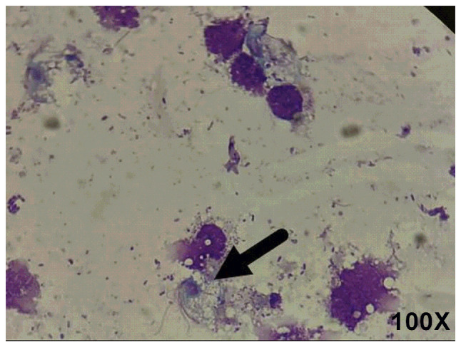
Giemsa staining. Samples were examined via light microscopy with Giemsa staining. The arrows indicate that a pear-shaped body with flagella was visible, containing a large dark purple long oval nucleus shaped like a mouse eye. There are four anterior flagella, and one posterior flagellum is as long as the undulatory membrane.