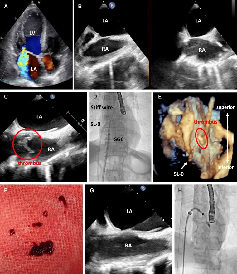
Figure 1.
(A) Transthoracic echocardiography on heart failure admission showed significant mitral regurgitation. (B) Transoesophageal echocardiography (TEE) with x-plane imaging before advancing steerable guide catheter (SGC) to right atrium (RA) revealed no structure in RA. (C) TEE immediately after advancing SGC to RA demonstrated a floating abnormal structure in RA. (D, E) Fluoroscopic imaging and 3D TEE during the structure aspiration in RA, respectively. (F) The aspirated structure was pathologically proven as thrombus later. (G) TEE after thrombus aspiration showed no structure in RA. (H) Fluoroscopic imaging after MitraClip implantation. Red circle indicates the thrombus in RA.

