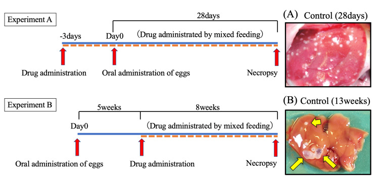
Figure 1. In vivo experiment protocol.
Overview of the experimental flow of the anti-echinococcal effect of drug candidates using mice with primary E. multilocularis infection. BALB/c mice were orally administered 300 eggs prepared from the feces of an E. multilocularis-infected dog (Experiment A, n = 10; Experiment B, n = 5). The images show typical cysts observed in an untreated mouse liver. (A) Cysts appear as small white dots on the liver. (B) Cysts are indicated by arrows.
E. multilocularis, Echinococcus multilocularis

