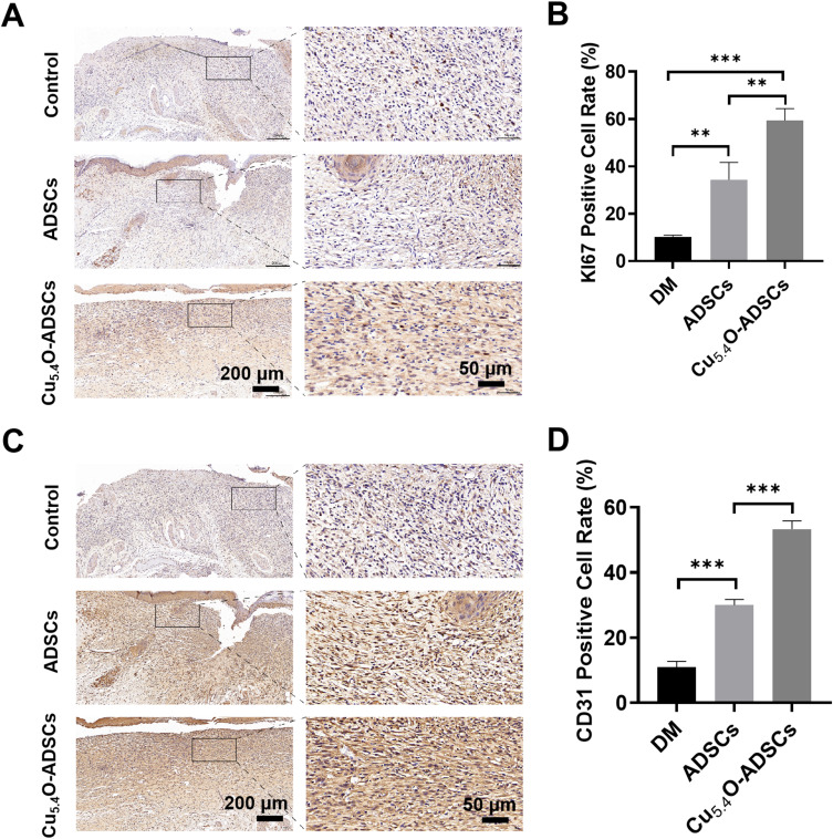
Figure 6.
(A) Immunohistochemical staining of Ki67 in wound tissues of diabetic mice from different treatment groups on postoperative day 14. (B) Quantitative analysis of Ki67 positive expression areas in diabetic mice wounds under different treatment conditions. (C) Immunohistochemical staining of CD31 in wound tissues of diabetic mice from different treatment groups on postoperative day 14. (D) Quantitative analysis of CD31 positive expression areas in diabetic mice wounds under different treatment conditions. ** P<0.01,***P<0.001.

