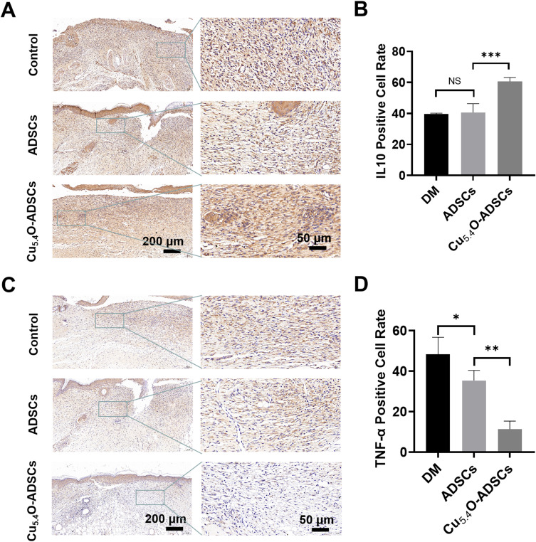
Figure 7.
(A) Immunohistochemical staining of IL-10 in wound tissues of diabetic mice from different treatment groups on postoperative day 14. (B) Quantitative analysis of IL-10 positive expression areas in diabetic mice wounds under different treatment conditions. (C) Immunohistochemical staining of TNF-α in wound tissues of diabetic mice from different treatment groups on postoperative day 14. (D) Quantitative analysis of TNF-α positive expression areas in diabetic mice wounds under different treatment conditions. *P<0.05,** P<0.01,***P<0.001, NS P>0.05.

