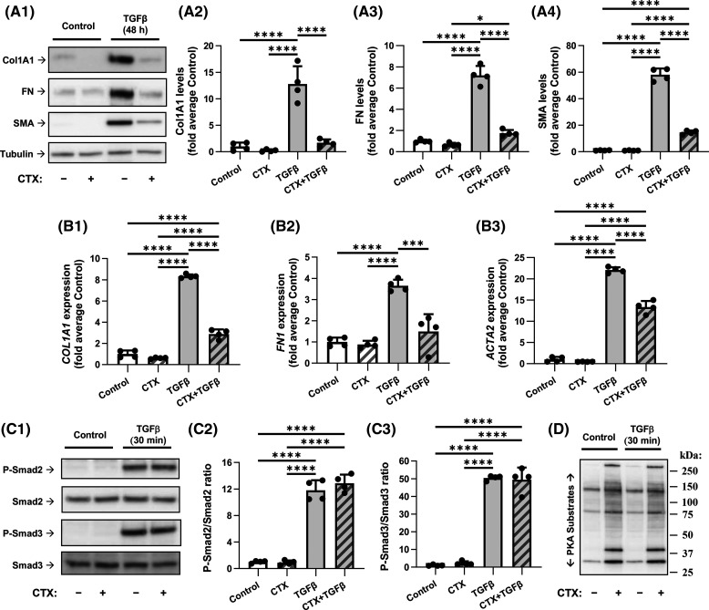
Figure 1. Activation of Gαs by cholera toxin blocks TGF-β-induced myofibroblast differentiation without affecting Smad2/3 phosphorylation.
(A) Representative images and quantification of western blot analyses of myofibroblast markers in human lung fibroblasts (HLF). HLF were serum-starved for 48 h and then treated with vehicle or 1 µg/ml cholera toxin (CTX), immediately followed by treatment with vehicle or 1 ng/ml TGF-β for additional 48 h, as indicated. HFL lysates were analyzed by western blotting using antibodies recognizing collagen 1A1 (Col1A1, A2), fibronectin (FN, A3) and smooth muscle α-actin (SMA, A4). The relative immunoreactivity values were normalized to the average signal of control samples. (B) RT-qPCR analysis of myofibroblast markers in HLF treated with vehicle, 1 µg/ml cholera toxin (CTX), and/or 1 ng/ml TGF-β (as in A) for 24 h. mRNA levels of COL1A1 (B1), FN1 (B2), and ACTA2 (B3) were normalized within-the-sample to the levels of housekeeping ribosomal RPL13 mRNA and compared with an average expression in control samples. (C) Representative images and quantification of western blot analyses of Smad2/3 phosphorylation in HLF pretreated with vehicle or 1 µg/ml CTX for 2 h followed by 30-min exposure to vehicle or 1 ng/ml TGF-β. Cell lysates were analyzed by western blotting with antibodies recognizing Smad2/3 and their phosphorylated forms as indicated. pSmad/Smad ratios were quantified for Smad2 (C2) and Smad3 (C3). (D) Western blot analysis of PKA-dependent phosphorylation in HFL treated as in C. and then probed with antibody recognizing phosphorylated PKA substrates (representative of three experiments). Quantitation data in A–C are the mean values ± SD of four independent treatments per group. *P < 0.05, ***P < 0.001, ****P < 0.0001, one-way ANOVA with Tukey correction for multiple comparisons.

