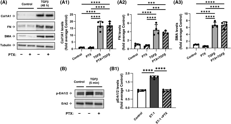
Figure 2. Inhibition of Gαi by pertussis toxin does not affect the TGF-β-induced myofibroblast differentiation.
(A) Representative images and quantification of western blot analyses of HLF pretreated overnight with 100 ng/ml pertussis toxin (PTX), followed by treatment with either vehicle or 1 ng/ml TGF-β for 48 h. HLF lysates were analyzed using antibodies recognizing Col1A1 (A1), FN (A2), and SMA (A3). The relative luminescence values were normalized to the average values of controls. Data are the mean values ± SD from four independent cultures per treatment. ***P < 0.001; ****P < 0.001, one-way ANOVA with Tukey correction for multiple comparisons. (B) Representative images of western blot analyses of the PTX pretreated HFL with or without subsequent 5-min treatment with 100 nM endothelin-1 (ET1). Cell lysates were probed with antibodies recognizing p-Erk1/2 or total Erk2. (B1) Quantification of experiments presented in B. ****P < 0.001, one-way ANOVA with Tukey correction for multiple comparisons.

