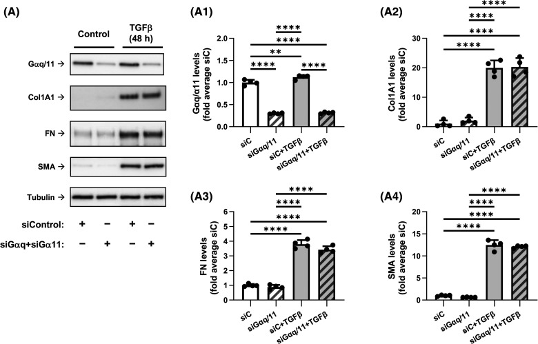
Figure 3. Knockdown of Gαq and Gα11 does not affect the TGF-β-induced myofibroblast differentiation.
(A) Representative images and quantification of western blot analyses of HLF transfected overnight with either control siRNA (siC) or with siRNAs targeting Gαq and Gα11. HFL were next serum starved for 48 h, followed by the treatment with either vehicle or 1 ng/ml TGF-β for 48 h. Cell lysates were analyzed using antibodies recognizing Gαq/Gα11 (A1), Col1A1 (A2), FN (A3), and SMA (A4). The relative luminescence values were normalized to the average siC-treated control samples. Data are the mean values ± SD from four independent cultures per treatment. **P < 0.01; ****P < 0.001, one-way ANOVA with Tukey correction for multiple comparisons.

