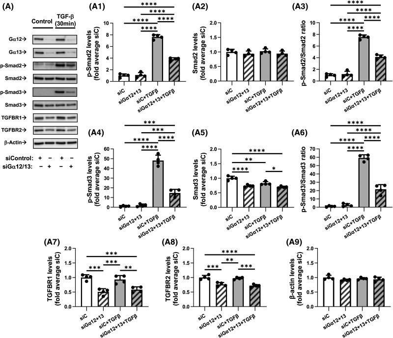
Figure 5. Combined knockdown of Gα12 and Gα13 inhibits TGF-β-induced Smad2/3 phosphorylation and reduces TGFβ receptor levels.
(A) Representative images and quantification of western blot analyses of HLF transfected overnight with control siRNA (siC, 10 nM) or with a combination of siRNAs targeting Gα12 (5 nM) and Gα13 (5 nM). HLF were next serum starved for 48 h and additionally treated with either vehicle or 1 ng/ml TGF-β for 30 min. Protein lysates were analyzed by western blotting using antibodies against p-Smad2, Smad2, p-Smad3, Smad3, TGFBR1, TGFBR2, or β-actin. Quantifications show chemiluminescence levels of p-Smad2 (A1), Smad2 (A2), ratios of p-Smad2/Smad2 (A3), p-Smad3 (A4), Smad3 (A5), ratios of p-Smad3/Smad3 (A6), TGFBR1 (A7), TGFBR2 (A8) and β-actin (A9), normalized to actin levels and average of siC. Data are the mean values ± SD from four independent cultures per treatment. *P < 0.05; **P < 0.01; ***P < 0.001; ****P < 0.001, one-way ANOVA with Tukey correction for multiple comparisons.

