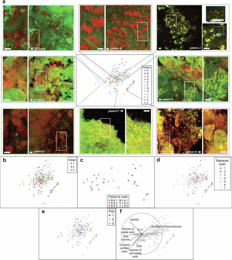
Fig. 3. Biofilm structure profiles generated by confocal microscopy.
Confocal laser scanning microscopy (CLSM) was performed for biofilms stained with a life/dead stain. a Non-metric Multi-Dimensional Scaling (nm-MDS) of 180 biofilm structure profiles captured from twelve patients, three time points, and five biofilm areas. Seven parameters –biofilm volume, red (dead) cell volume, green (live) cell volume, percent of live cell volume, number and size of microcolonies, and surface area covered by biofilm – were standardized by maximum and fourth root transformation prior to pairwise calculations of Euclidean distances. Symbols of different shape and color indicate different patients. Eight examples of confocal images are presented. Bar indicates 40 µm. b Symbols indicate age of the biofilms. c Enlarged nm-MDS of biofilm structure profiles of two selected patients. 2D-stress was 0.06. d Symbols indicate implant location. e Symbols indicate the Periodontal Screening Index (PSI). f Superimposed is a vector plot for biofilm (in grey) and clinical (in red) parameters, with the vector direction for each class reflecting the Pearson correlations of their values with the ordination axes, and length giving the multiple correlation coefficient from this linear regression on the ordinate points.

