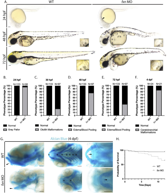
FIGURE 2.
Morphological analysis of fxn-deficient zebrafish embryos reveal phenotypes indicative of several developmental malformations. (A) Brightfield microscopy revealed a diffuse gray pallor in the cranium of 24 hpf fxn morphants as well as blood pooling and pericardial edema at the 48 and 72 hpf stages. Insets show otolith malformations in the fxn morphants. Scale bars = 50 um. (B–F) Penetrance of observed phenotypes. (B) Gray pallor at 24 hpf. (C) Otolith malformations at 30 hpf. Edema/blood pooling at (D) 48 hpf and (E) 72 hpf. (F) Ceratobranchial malformations at 4 dpf. (G) Alcian blue staining revealed ceratobranchials 1-5 were missing in fxn morphants. Additionally, Meckel’s cartilage failed to fuse and the mandibular prominence did not curve ventrally as it does in wild-type animals. Scale bars = 50 um. (H) Kaplan-Meier survival curve documenting survival in wild-type embryos compared to fxn morphants. Fxn morphant animals displayed a 64% chance of survival compared to the 94% chance of their wild-type siblings during the first 12 days of life.

