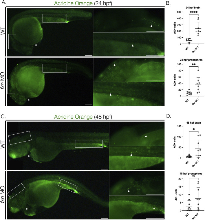
FIGURE 7.
Cell death is elevated in fxn-deficient zebrafish embryos. (A) Images of AO-stained animals at 24 hpf. Fxn morphants animals have significantly increased cell death in the brain and pronephros at 24 hpf and increased cell death in the brain at 48 hpf. White boxes denote the locations for the panels on the right top (brain) and right bottom (pronephros). Scale bars = 50 um. (B) Unpaired t-tests of cell death in the brain and pronephros at 24 hpf. (C) Images of AO-stained animals at 48 hpf. White boxes denote the locations for the panels on the right top (brain) and right bottom (pronephros). Within the right top brain panel, each white box indicates a single positive cell. In fxn morphants, areas of the brain commonly had multiple cells as indicated by the white box. Scale bars = 50 um. (D) Unpaired t-tests of cell death in the brain and pronephros at 48 hpf. *p < 0.05, **p < 0.005, ****p < 0.0001.

