. 2024 Oct 15;17(4):383–388. doi: 10.3400/avd.oa.24-00031

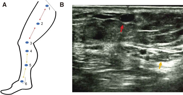
Fig. 1 (A) Scheme and the definition of the 6 points. Point 1: The sapheno-femoral junction was depicted. Point 2: The midpoint between points 1 and 3. Point 3: The knee joint level. Point 4: The saphenous nerve and the GSV could be seen on the ultrasonography monitor simultaneously below the knee (B). Point 5 was the midpoint between points 4 and 6. Point 6: The level of the medial malleolus. Straight dotted line: inguinal ligament, curved dotted line: medial malleolus. The distances between 1 and 2, 2 and 3 are equal (red arrowed line). The distances between 4 and 5, 5 and 6 are equal (yellow arrowed line). (B) The GSV and the saphenous nerve at the point 4. Red arrow: GSV; yellow arrow: saphenous nerve. These 2 components are depicted simultaneously. In this case, the distance between them is 14 mm. GSV: great saphenous vein.

Fig. 1 (A) Scheme and the definition of the 6 points. Point 1: The sapheno-femoral junction was depicted. Point 2: The midpoint between points 1 and 3. Point 3: The knee joint level. Point 4: The saphenous nerve and the GSV could be seen on the ultrasonography monitor simultaneously below the knee (B). Point 5 was the midpoint between points 4 and 6. Point 6: The level of the medial malleolus. Straight dotted line: inguinal ligament, curved dotted line: medial malleolus. The distances between 1 and 2, 2 and 3 are equal (red arrowed line). The distances between 4 and 5, 5 and 6 are equal (yellow arrowed line). (B) The GSV and the saphenous nerve at the point 4. Red arrow: GSV; yellow arrow: saphenous nerve. These 2 components are depicted simultaneously. In this case, the distance between them is 14 mm. GSV: great saphenous vein