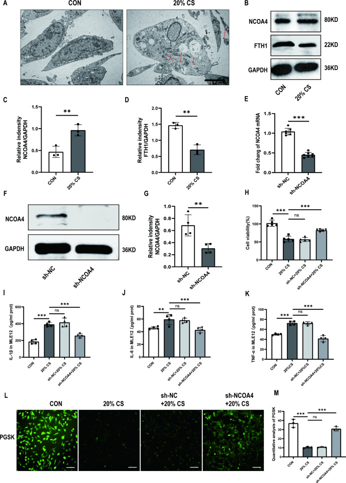
Fig. 2.
Ferritinophagy promotes ventilator-induced ferroptosis of MLE-12 cells through ferritin degradation mediated by NCOA4. (A) Representative TEM images of MLE12 cells untreated or treated with 20% CS. Red arrows represent autophagosomes. Magnifications: 5000 X, acceleration voltage: 80 kV. Scale bar: 5.0 μm. (B) Representative Western blotting bands of NCOA4, FTH1, and GAPDH in MLE12 cells. (C, D) Relative protein expression of NCOA4 and FTH1 was divided into GAPDH (n = 3). (E) The mRNA levels of NCOA4 knockdown in MLE12 cells (n = 7). (F) Representative Western blotting images of NCOA4 and GAPDH in MLE12 cells. (G) The protein expression of NCOA4 relative to GAPDH (n = 4). (H) Cell viability was detected using a CCK8 assay (n = 5). (I-K) Levels of IL-1β, IL-6, and TNF-α in MLE12 cells (n = 4). (L) PGSK probe staining for Fe2+ in MLE12 cells with different treated groups. Scale bar: 100 μm. (M) Quantification of the fluorescence intensity of PGSK using ImageJ Fiji software (n = 3). Data are expressed as mean ± SD. “*” indicates significant differences between groups (*p < 0.05, ** p < 0.01 or *** p < 0.001)

