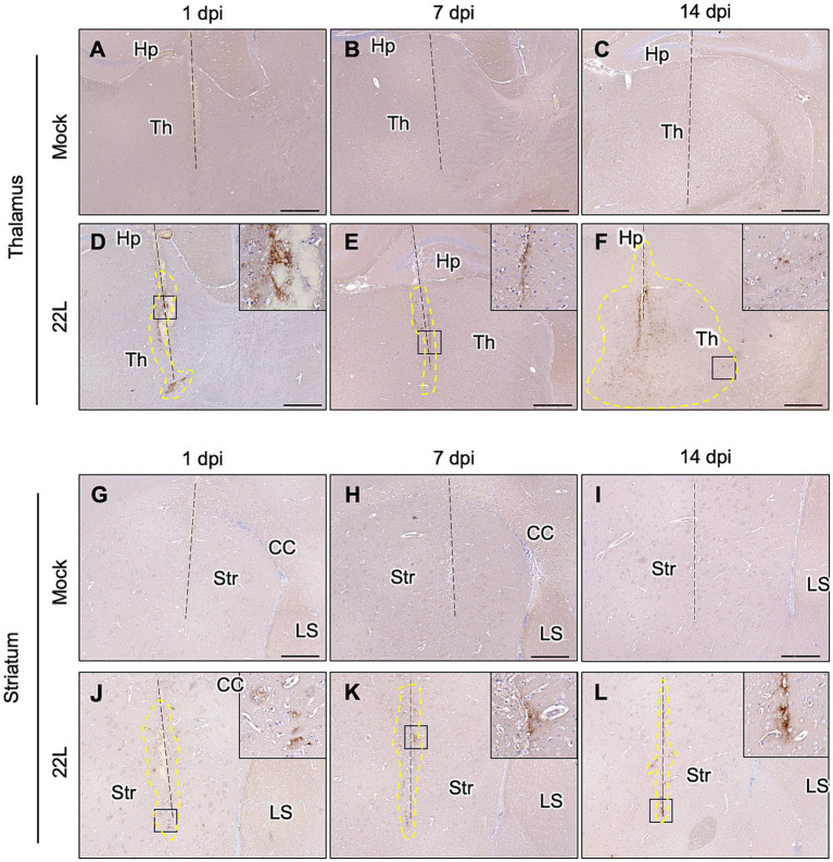
Figure 2.
PrPSc detection after stereotaxic injection of prions. Brain homogenates from mock- or 22L prion-infected mice (0.1%, 0.5 μL) were injected into the thalamus (A–F) or striatum (G–L) using a stereotaxic apparatus. Brains were collected at 1 (A,D,G,J), 7 (B,E,H,K), and 14 (C,F,I,L) days post-injection and fixed with 10% neutral buffered formalin. Coronal sections corresponding to Plates 45–46 (thalamus) and 22–23 (striatum) (Paxinos and Franklin, 2013) were cut for immunohistochemical detection of PrPSc using anti-PrP mAb 132. FFPE blocks containing either the thalamus or striatum were serially cut into 4 μm sections at the coronal plane using a microtome. Sections in which needle tracks were observed were used for PrPSc staining with IHC. Dashed lines indicate needle tracks. Higher magnifications of boxed regions in the 22L strain-injected thalamus and striatum are shown in the upper-right corners of each lower magnification image. The region surrounded by dotted lines in the 22 L strain-injected thalamus (F) indicates the spread of PrPSc-positive regions from the needle track. Th, thalamus; Hp, hippocampus; Str, striatum; CC, corpus callosum; LS, lateral septum. Scale bars: 300 μm. Dotted lines indicate regions with positive signals determined using ImageJ (ver 1.8.0).

