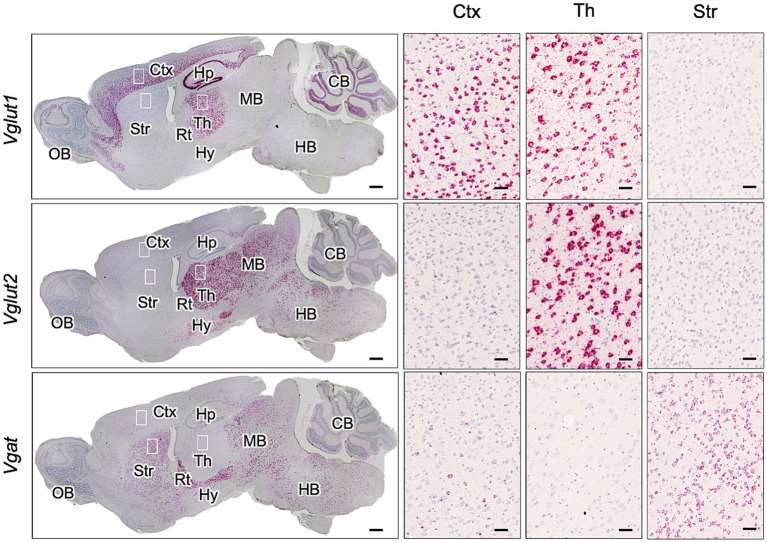
Figure 3.
Expression of Vglut1, Vglut2, and Vgat mRNA in mouse brain. RNAscope in situ hybridization was used to detect the gene expression of Vglut1 and Vglut2, markers for glutamatergic neurons, and Vgat, a marker for GABAergic neurons, in sagittal brain sections from an uninfected adult ICR mouse. The leftmost column displays a whole sagittal section, while the three images on the right show magnified views of the boxed regions in the cortex (Ctx), thalamus (Th), and striatum (Str). Positive signals for the target RNA appear reddish, developed with RNAscope® 2.5 HD Detection Reagent (RED), while counterstaining with hematoxylin produces light blue staining. Images were captured with a NanoZoomer 2.0RS (Hamamatsu Photonic K.K., Hamamatsu, Japan) using a 40× objective and stitched with NanoZoomer 2.0RS software (Hamamatsu Photonic K.K., Hamamatsu, Japan). Scale bars are 1 mm in the sagittal plane images and 0.1 μm in the magnified images. OB, olfactory bulb; Hp, hippocampus; Rt, reticular nucleus; Hy, hypothalamus; MB, midbrain; HB, hindbrain; CB, cerebellum.

