Figure 8.
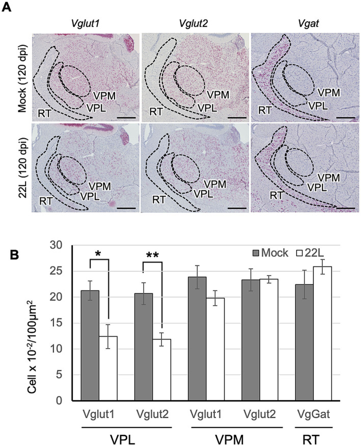
Type of thalamic neurons lost in prion infection. (A) RNAscope analysis: RNAscope in situ hybridization was used to detect the gene expression of Vglut1 and Vglut2, markers for glutamatergic neurons, and Vgat, a marker for GABAergic neurons, in the thalamus of prion 22L strain-infected mice at 120 dpi. Coronal sections around Plate 45–46 (Paxinos and Franklin, 2013) were used. Positive signals for the target RNA were developed with RNAscope® 2.5 HD Detection Reagent—RED, showing a reddish color, while counterstaining with hematoxylin produced light blue staining. Images were captured with a NanoZoomer 2.0RS using a 40× objective and stitched with NanoZoomer 2.0RS software. The ventral posterolateral nucleus (VPL), ventral posteromedial nucleus (VPM), and reticular nucleus (RT) are enclosed with dashed lines. Scale bar: 0.5 mm. (B) Quantitative analysis: Neurons positive for Vglut1, Vglut2, or Vgat were counted as described in the materials and methods. Graphs show the number of positive cells per 100 μm2 (mean ± SD of 3 brains from either mock- or prion-infected mice). Three sections were analyzed from each mouse. Statistical analysis was performed using Student’s t-test, *p < 0.05, **p < 0.01.

