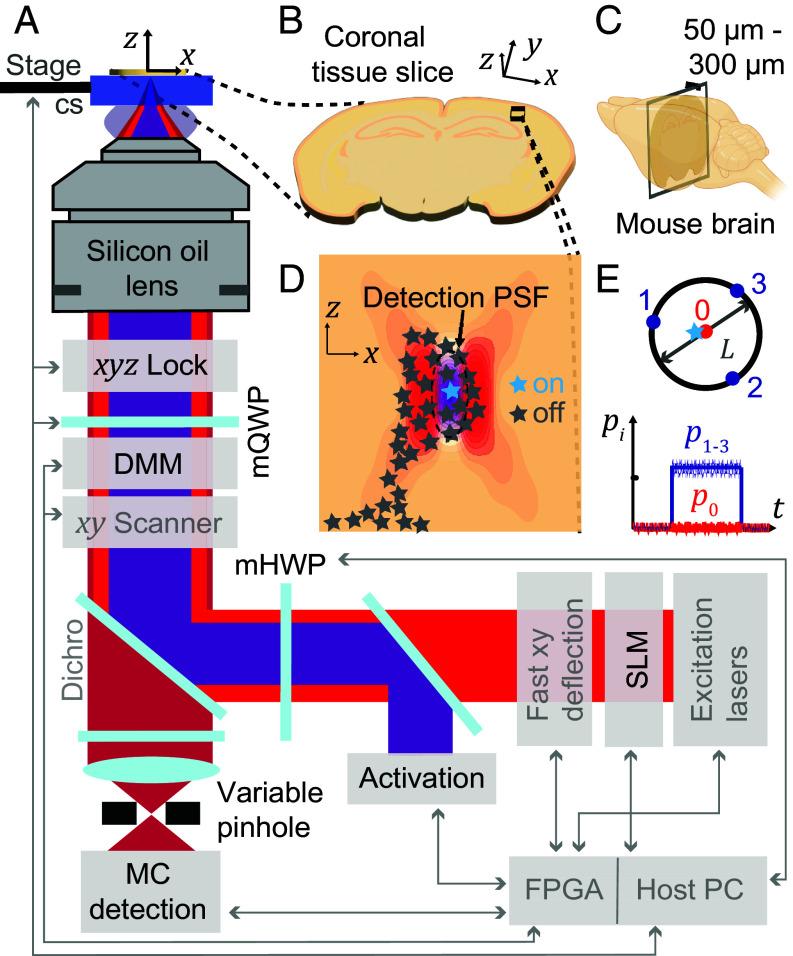
Fig. 1.
MINFLUX nanoscopy in biological tissue. (A) Overview of the custom-built MINFLUX setup for imaging in tissue (adaptations for tissue imaging highlighted in black, standard MINFLUX components shown in gray). (B) Sample imaged. The sample is a coronal section prepared by slicing (C) a mouse brain into 50-300 micrometer consecutive sections. (D) Region of interest in the brain slice showing the optical sectioning capabilities of MINFLUX by the confocal detection volume and the selective on- and off-switching of single emitters. The activated fluorophore is localized photon-efficiently by centering the zero of the donut excitation beam on the activated molecule. (E) Targeted-coordinate pattern for the donut excitation beam and schematic photon emission trace of a centered molecule. cs: coverslip; DMM: deformable membrane mirror; mQWP, mHWP: motorized quarter/half-wave plates; pi: photon count ratio in ith exposure; MC: multicolor; SLM: spatial light modulator; FPGA: Field-Programmable Gate Array.

