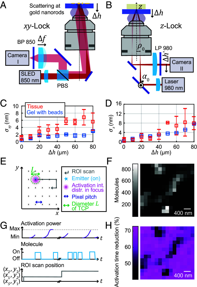
Fig. 2.
Active drift-correction for depth imaging and progressive activation. (A) Depth-adaptable xy-lock system. The dark-field images of fiducial markers (gold nanorods) on the coverslip are acquired when imaging in a plane away from the coverslip surface by refocusing the nanorods with a variable-focus lens. (B) Depth-adaptable z-lock system. The imaging depth range that can be stabilized with high precision is enlarged by a piezo-actuated closed-loop absolute positionable mirror mount. (C and D) Stability of the (C) xy-lock position and (D) z-lock position for measurements in both agarose-sucrose and tissue samples (mean stabilities ± SD). (E) Schematic of activation scan over the ROI. (F) Number of fluorophores detected at each mosaic scan position. (G) Progressive activation, shown schematically. At each new activation location addressed by the mosaic scan, the activation power is ramped up until a molecule starts to emit. (H) Reduction in activation time for the progressive activation relative to activation time expected with constant low activation. PBS: polarizing beam splitter, BP, LP: band/long pass, Δh: imaging depth, α0: rotation angle of absolute positionable mirror, Δh’: z-lock beam position on camera, σxy, σz: xy-lock and z-lock position stability, SLED: superluminescent diode.

