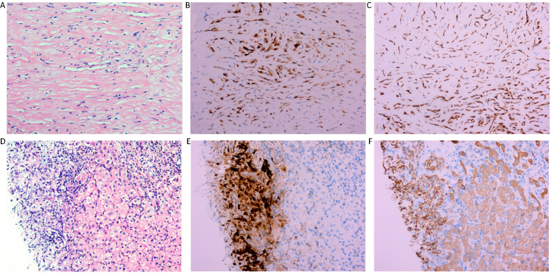
Figure 4.
Histological findings of the right subdiaphragmatic lesion. Hematoxylin-eosin (HE) staining (x 200) reveals dense fibrocollagenous tissue infiltrated by spindle-shaped cells (A). and immunohistochemistry revealed immunopositivity for CR and CK (B, C), suggestive of desmoplastic malignant mesothelioma of peritoneum. Histological findings of the lesions of the liver. Hematoxylin-eosin (HE) staining (x 200) reveals dense fibrocollagenous tissue infiltrated by spindle-shaped cells (D) and immunohistochemistry showing immunopositivity for CR and CK (E, F), which is indicative of desmoplastic malignant mesothelioma of the peritoneum directedly invading the liver.

