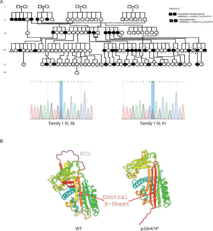
Fig 1.
SERPING1 variant on Family 1 A) Pedigree of Family 1 with persons harboring the variant c.1420C>T p.(Gln474*) and Sanger sequencing confirmation of IV,38 and IV,41.+WES and *or ** negative or positive SS respectively. B) Left, the wild-type SERPING1 protein structure with the reactive center loop (RCL) highlighted in magenta. The central beta-sheet region is indicated in red; right, SERPING1 protein structure featuring the Gln474* variant. The molecular variant promotes the insertion of the RCL into the central beta-sheet, mimicking the latent form of the protein. The absence of the RCL region is shown, and the central beta-sheet is indicated in red. The overall conformational change depicts the predicted structural transition associated with the pathogenic variant.

