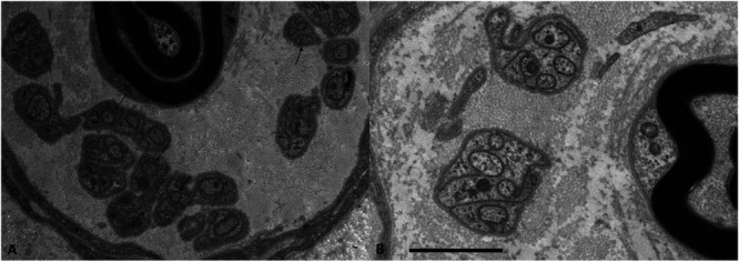
Figure 1.

Example of axons with reduced diameters in patients with FMS. Electron micrograph of a Remak bundle. Numerous unmyelinated fibers of small diameter (arrows) are found in a patient with FMS (A), but not in a normal control (B). Bar = 2 mm. FMS, fibromyalgia syndrome. Figure from Reference 15, RightsLink licence number 5817811230620.
