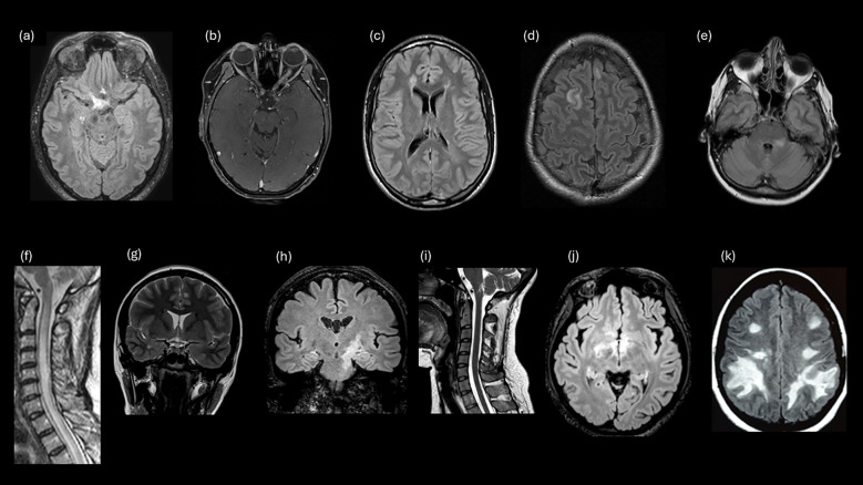
Figure 1.
Examples of study survey images that evaluated participant knowledge for MRI lesions. (A) Lesion involving optic chiasm and posterior ON. (B) LEON (longitudinally extensive left optic neuritis). (C) Periventricular (MS). (D) Juxtacortical (MS). (E) Fluffy lesion and poorly demarcated lesion (MOGAD). (F) LETM. (G) Cortical lesion lob temporal (MS). (H) Corticospinal tract lesion. (I) Lesion involving the area postrema. (J) Hypothalamic lesion. (K) Large, confluent, bilateral subcortical or deep white matter lesions.

