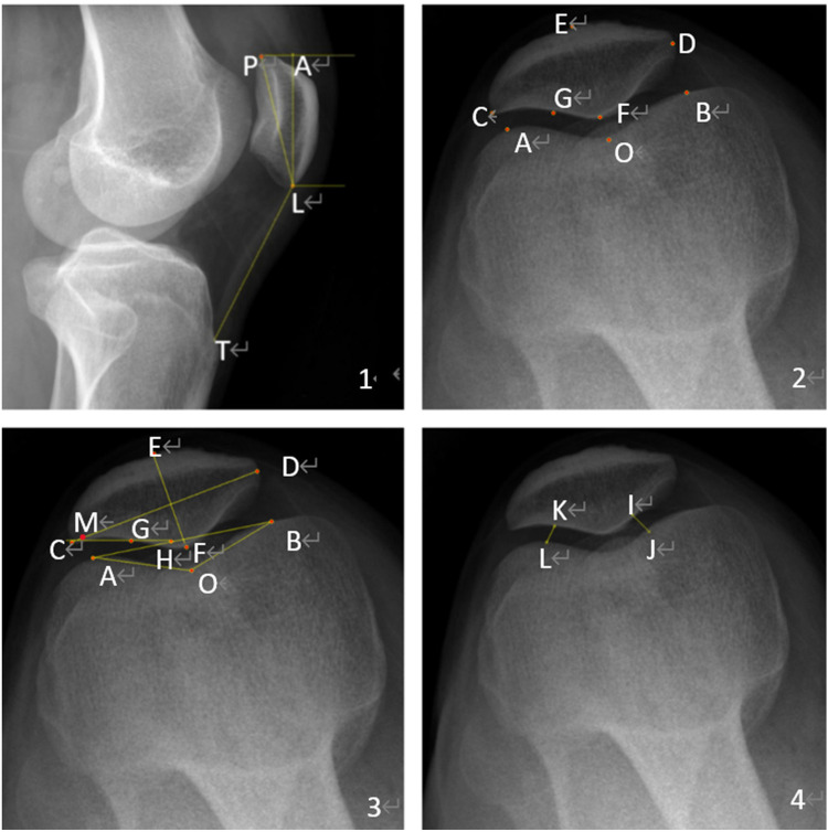
Figure 2.
Measurement parameters for the patellofemoral. 1. X-ray of the lateral patellofemoral. 2. Location of landmarks on an axial x-ray image. P superior pole of the patella. L Inferior pole of patella. T Tibial tuberosity inferior pole. The Insall-Salvati ratio = LT/LP, and the patellar length = AL (Figure 2.1). The highest point of the lateral condyle. B Highest point of the medial condyle. C Outermost point of patella. D Most medial point of patella. E Anterior cortex of the patella. F Median cartilaginous ridge of patella. G Tangent point of the lateral articular surface of the patella (Figure 2.2). H The intersection of the lateral patella's articular surface and the femoral internal and external condyles line. M is the point at which the highest point of the lateral epicondyle of the femur intersects with the width of the patella. O Most distal point at the bottom of the trochlear groove Patellar width = CD, Patella thickness = EF. Groove angle = angle AOB. Patellar displacement rate = CM/CD. Lateral patellofemoral angle = angle GHA (Figure 2.3). IJ, KL, the patellofemoral joint's internal and lateral spaces, are connected in the shortest distance. Patellofemoral index = IJ/KL (Figure 2.4).

