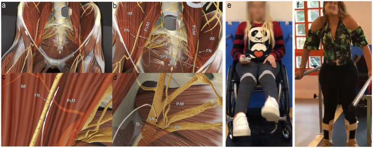
Figure 1.
Electrodes placement and nerve stimulation. (A) Panoramic view of the system; the pulse generator is implanted into a paraumbilical subcutaneous pocket, and the electrodes run retroperitoneally down to the intrapelvic portions of the femoral (FN), sciatic (SN), and pudendal nerves (PN) bilaterally. (B) Panoramic view of femoral electrodes (FEs) and sciatic and pudendal electrodes (SPEs) positioning. (C) Detailed view of the right femoral electrode over the nerve between the iliac (IM) and the psoas (PsM) muscles. (D) Detailed view of the right sacral electrodes placed with half of its poles in the Alcock’s canal over the PN and the remaining poles over the SN. IS, ischial spine; PiM, piriformis muscle. (E) Extension of knees through femoral nerve stimulation using a remote activation. (F) Weight transfer and walking on parallel bars. Figure adapted from Lemos et al. (2023), with permission from the authors.

