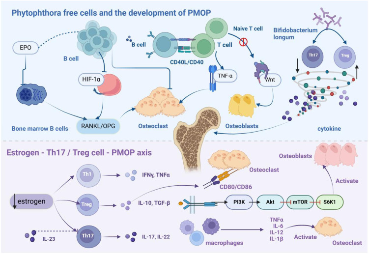
Figure 1.
Immunopathological Mechanism of PMOP. B cells differentiate into osteoclasts, leading to bone loss. In the estrogen-deficient state, hypoxia-inducible factor-1α signaling is activated in B cells, enhancing RANKL expression and promoting osteoclastogenesis, thereby inducing PMOP. T cells normally exhibit bone-protective functions in basal bone metabolism. However, following ovariectomy, CD4+ and CD8+ T cells become activated and secrete RANKL and other osteoclastic factors. Treg cells secrete IL-10, TGF-β, and other anti-resorptive cytokines, whereas Th17 cells produce IL-17, which stimulates osteoclastogenesis. Additionally, IL-17 triggers mesenchymal stem cells to release osteoclast differentiation factors. Thus, estrogen modulates bone metabolism by regulating the balance between Th17 and Treg cells.

