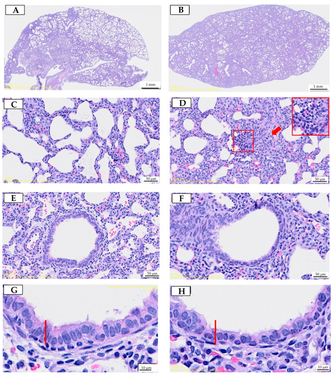
Figure 1.
Prenatal polystyrene microplastics (PS-MPs) exposure leads to lung dysplasia in offspring during infancy. Histological manifestations of lung tissue from seven-day-old offspring with maternal microplastic particle exposure are shown. (A) Control group: under low-power microscopic field, the control group exhibited better pulmonary tissue aeration development. (B) PS-MPs exposure (1000 µg/L in drinking water for pregnant dams). (C) Zoomed-in images of Figure 1A. (D) Zoomed-in images of Figure 1B, showing the collapsed alveoli and thickened alveolar septa (red arrow). Magnification of boxed area detailing the inflammatory cells; cross-section of small airway from the (E) control group and (F) prenatal PS-MPs exposure group. (G) Zoomed-in images of (E). (H) Zoomed-in images of (F). The columnar epithelial cells in the airway of the prenatal PS-MPs exposure group are shorter and less developed, as indicated by the red marker representing the height of the control group cells. Each group comprised six animals. The yellow label at the bottom left of (F) and the top right of (G) is a specimen group annotation, with no other significance.

