Figure 5.
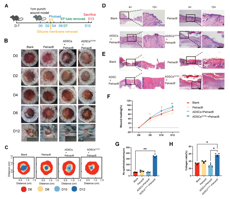
Dermal substitute–ADSCsDGTM+ complexes fill full-thickness defective wounds. (A) Flowchart of animal experiments. (B) Representative images of skin regeneration. Mice wounds were divided into the following groups: blank, Pelnac® ADSCs + Pelnac®, and ADSCsDGTM+ + Pelnac®. (C) Temporal variation of skin regeneration. (D) HE staining of mice in each group of the wound. (E) MASSON staining of mice in each group of the wound. (F) Statistical graph of the percentage of wound healing over time for each group. (G) Statistical graph of the thickness of re-epithelialization of the wound in each group. (H) Statistical graph of the proportion of wound collagen in each group. * p < 0.05, ** p < 0.01, n = 4.

