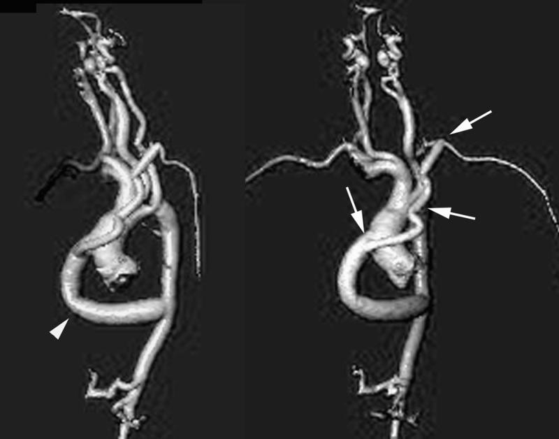
Fig. 3 Postoperative, contrast-enhanced, 3-dimensional, magnetic resonance angiograms. A) The lateral view reveals severe hypoplasia of the transverse arch, as in Figure 1. However, the patient now has 2 grafts: the larger, 16-mm graft (arrowhead) is anastomosed end-to-side to the mid-portion of the ascending aorta and to the distal portion of the descending thoracic aorta. B) The shallow left anterior oblique projection shows the smaller, 7-mm graft (arrows), which has been sutured end-to-side to the proximal 16-mm graft and to the left subclavian artery.
