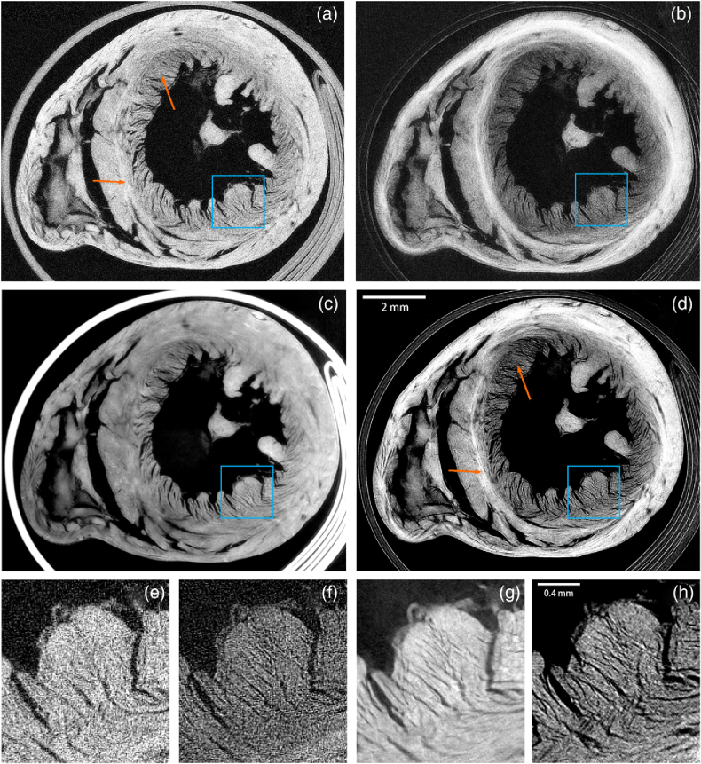
Fig. 4.
Tomographic slices of dried rat heart sample shown with (a)+(e) attenuation contrast, (b)+(f) dark-field contrast, (c)+(g) phase-contrast, and (d)+(h) hybrid contrast with Eq. (14) and . All slices from datasets with matched total exposure time, with attenuation, phase, and dark field from a fully sampled dataset. Magnified sections in the blue ROI are shown below for the corresponding slices in (e)–(h). The hybrid contrast retains area contrast in the wall (orange arrows) from the dark-field slice, not present in the attenuation slice. Additionally, the inserts show much more detail with hybrid contrast than attenuation contrast.

